Foot and Ankle Surgeon Helps With Foot Deformities
Foot and Ankle Surgeon in Shasta County Helps People Recover from Foot Deformities
Dr. Garret Strand, DPM
Dr. Garret Strand, DPM, is one of Shasta Orthopaedics Foot & Ankle surgeons who provides people with reconstructive surgery for foot deformities. He has extensive experience in trauma, elective reconstruction, arthroscopy, and total ankle replacement surgery.
This article shares four case studies, with photos of reconstructive surgery, of patients who came to Shasta Orthopaedics for treatment of various foot deformities.
What is a Foot Deformity?
Foot deformities mean that something about the shape of the foot is not typical. Some people are born with foot deformities. For example, 1 in every 1,000 babies in the United States are born with clubfoot deformity, where the feet are rotated inward and downward. Not all foot deformities cause people pain or need reconstructive surgery. Non-invasive procedures and over-the-counter medication can help with some foot deformities. Shasta ortho provides non-surgical foot and ankle treatments, when appropriate.
In other cases, reconstructive surgery is necessary to alleviate debilitating pain caused by shifted or damaged bones, joints, muscles, ligaments, or tendons. Shasta Orthopaedics is dedicated to the prevention, diagnosis, and treatment of these conditions. As a foot doctor or DPM, Dr. Strand began sharing posts on Instagram @shastaorthofootandankle to address surgeons’ and patients’ questions about reconstructive foot and ankle surgery.
Below are four case studies highlighting recent reconstructive surgeries.
Case Study 1 – Reconstructive Surgery for Patient with Polymetatarsia
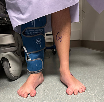
Reconstructive Surgery for Patient with Polymetatarsia
This patient came to Shasta Orthopaedics with a history of polymetatarsia, which is a malformation in the foot caused by having six or more metatarsal bones in the same foot. Years prior, this patient had the polymetatarsia surgically removed. As a fourteen-year-old, they expressed having difficulty participating in physical activity, decreasing weight bearing (WB) tolerance, and difficulty finding comfortable shoe gear.
The patient also had magnetic resonance imaging (MRI) with partial fusion to their first tarsometatarsal (TMT). The patient underwent Gastrocnemius recession, Evans calcaneal osteotomy, first TMT medial, plantar closing wedge arthrodesis, 1st and 2nd metatarsal-phalangeal joint (MTP) capsulotomies, and 1st MTP rebalancing with forefoot box stitch internal brace.
Now, the patient can participate in physical activity for increased periods of time and also wear shoe gear more comfortably. When the patient is ready, Dr. Strand will do the same with the other foot.
Case Study 2 – Reconstructive Surgery for Patient with Pes Planus Deformity
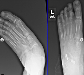
Reconstructive Surgery for Patient with Pes Planus Deformity
This patient came to Shasta Orthopaedics with severe pes planus deformity in their left foot. Pes planus is a foot deformity where the foot is closer to the ground due to the loss of the medial longitudinal arch in the foot.
At age 13, this patient exhibited pes planus deformity, equinus contracture, hindfoot with calcaneal valgus, forefoot abduction, and medial column collapse. They also had subtle limb-length discrepancies. With these additions, it was important to approach this surgery sequentially from proximally to distally.
First, the patient underwent Gastrocnemius recession followed by tendon Achilles lengthening (TAL) due to their residual equinus contracture, followed by arthroereisis; DPMs at Shasta Orthopaedics rarely use these implants but this patient was a strong candidate due to the severity of his deformity and laxity; Dr. Strand didn’t fully trust the amount of correction that bony and soft tissue would provide and the patient’s subtalar joint (STJ) had lots of subluxation. The DPMs needed to buttress or stop the amount of motion at this joint and given the patient’s age, he was a good candidate for arthroereisis.
Next, the patient underwent Evan’s calcaneal osteotomy, cotton medial cuneiform osteotomy, Kidner posterior tibial tendon advancement, and spring ligament repair. At six months out, the patient is comfortable and back in supportive shoes with inserts.
Case Study 3 – Reconstructive Surgery for Patient with Charcot Foot
Reconstructive Surgery for Patient with Charcot Foot
Charcot foot can develop as a result of broken bones or nerve damage, among other causes. Often, nerve damage causes a weakening of bones; when this happens, toes can turn inward or the ankle can become deformed.
At age 53, this patient had Charcot deformity in their right foot. Once out of the acute phase, they underwent initial Charcot reconstruction. Six months post-operation, the patient underwent another Charcot reconstruction and broke through the medial column plate. Computerized tomography (CT) confirmed the nonunion and fragmentation of multiple joints.
Because of this, the patient then underwent Charcot reconstruction with hardware removal (HWR) and beaming. After five months and s/p revision, the patient is back in supportive shoes and their daily activities.
Case Study 4 – Reconstructive Surgery for Patient with Charcot Foot
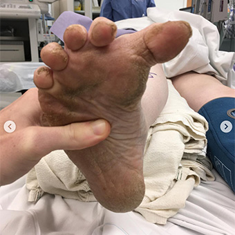
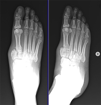
Reconstructive Surgery for Patient with Charcot Foot
At age 74, this patient expressed worsening right foot pain and Charcot deformity. The patient had undergone multiple prior procedures for this foot and had not been able to wear closed-toed shoes for nine years.
Clinical and radiographic evaluation showed large hallux varus deformity with hallux malleolus, global arthritic changes to the foot, and a collapsed medial longitudinal arch with a plantar flexed cuboid.
To address this, Dr. Strand elected for a 1st MTP arthrodesis with interphalangeal joints
(IPJ) arthrodesis, along with 2-5 Hammertoe (HT) correction and MTP capsulotomies.
One of the tricks to reducing and correcting this incredible deformity was shortening the 1st ray through our joint prep. This case shows the deformity correctional power of an excellent 1st MTP arthrodesis.
Early Diagnosis and Treatment Helps People with Foot Deformities
The above four cases show that people of any age who are experiencing pain from a foot deformity might be great candidates for reconstructive surgery. With all foot and ankle problems, early diagnosis and treatment help prevent further damage and complications.
If you have a foot or ankle problem, get it seen by a qualified foot and ankle specialist as soon as possible.
